Which Defect Results in Increased Pulmonary Blood Flow
These are all abnormal openings in the heart well talk more specifically about their locations in just a second. The pulmonary artery is narrowed so blood cant easily flow out of the right ventricle.

Pulmonary Valve Atresia An Overview Sciencedirect Topics
Lets start off by talking about defects that cause increased pulmonary blood flow.

. Defects with Increased Pulmonary Blood Flow. Up to 24 cash back This is the combination of four heart defects that interfere with the blood flow to the lungs. May be asymptomatic but with a.
There is a low-placed Atrial Septal Defect a high-placed Ventricular Septal Defect and fusion of the mitral and tricuspid valves allowing blood to flow between all heart chambers. - atrial septal defect asd an abnormal opening between the atria that causes an. Hypertrophy of the right ventricle due to pulmonary stenosis.
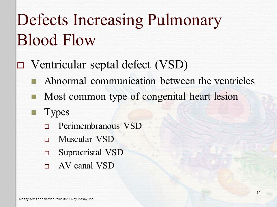
Ventricular septal defect VSD what happens to the size of the heart in atrial septal defect. The increases pressure on the right side and causes deoxygenated blood to shunt through the VSD to circulate through the body. The atrial septal defect results in increased pulmonary blood flow.
This includes an atrial septal defect or ASD a ventricular septal defect or VSD an AV canal defect as well as a patent ductus arteriosus or PDA defect. Neonates with congenital cardiac defects with increased pulmonary blood flow Abstract Generally cardiac lesions with increased pulmonary blood flow demonstrate cardiomegaly increased pulmonary vascular markings and pulmonary congestion on the chest x-ray. Results of treatment of patients with ventricular septal defects.
This causes an increased flow of oxygenated blood into the right side of the heart. What defect is an abnormal opening between atria. The truncus arteriosus overlies a VSD that is almost always seen in conjunction with this defect.
Left to right shunting occurs resulting in pulmonary vascular engorgement and HF. The muscle of the right ventricle is thick and overworked. Atrial septal defect Click card to see definition Defect in septum that leads to increased blood to the right side because the pressure on left is higher.
Ventricular Septal Defect VSD. In our model of a congenital heart defect CHD with increased pulmonary blood flow PBF. Intracardiac communication along the septum or an abnormal connection between the great arteries allows blood to flow from the high-pressure left side of the heart to the low-pressure right side of the heart.
The defects that cause an increase in pulmonary blood flow are Atrial Septal Defect Ventricular Septal Defect Atrioventricular Canal Defect and Patent Ductus Arteriosus. The four defects include. Pulmonic stenosis ventricular septal defect overriding aorta right ventricular hypertrophy.
An overriding aorta that is displaced to the right so there is flow from both ventricles. These are all abnormal openings in the heart well talk more specifically about their locations in just a second. Atrial septal defect ASD what defect is an abnormal opening between ventricles.
This article examines common cyanotic congenital heart lesions that result in an increase in pulmonary blood flow. Increased pulmonary blood flow at increased pulmonary artery pressure. Cyanotic congenital heart disease with increased pulmonary blood flow Abstract Pediatricians daily encounter children with systemic cyanosis.
Second natural history study of congenital heart defects. Leading to right-to-left shunting -Hypoxemia and cyanosis result from bc of the increased amount of unoxygenated blood in the systemic circulation. These data suggest that increased pulmonary blood flow from ventricular septal defect results in altered carnitine and mitochondrial homeostasis decreased nitric oxide signaling and increased reactive oxygen species production.
Tricuspid valve abnormalities include atresia hypoplasia ie pulmonary atresia with intact ventricular septum and displacement ie Ebstein anomaly. Defects with increased pulmonary blood flow. Which of the following defects results in increased pulmonary blood flow.
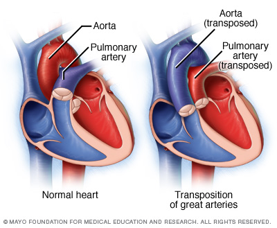
AnatomyTruncus arteriosus is a rare congenital heart defect in which a single great vessel arises from the heart giving rise to the coronary systemic and pulmonary arteries. Congenital heart defects may cause cyanosis. Transposition of the great arteries ANS.
Atrial septal defect 4. The defects that cause an increase in pulmonary blood flow are Atrial Septal Defect Ventricular Septal Defect Atrioventricular Canal Defect and Patent Ductus Arteriosus. Cyanotic Defects Mnemonic Hypoxia Signs and Symptoms in Pediatrics Mnemonic Congenital Heart Defects Care Plans Outline Overview Pulmonary blood flow is obstructed Causing pressure to be higher in the Rt not the Lt Deoxygenated blood is then shunted from the Rt side of heart to Lt side.
-decrease the amount of blood that gets oxygenated by the lungs -the obstructed pulmonary blood raises pressures on the hearts Right side higher than the Left. Pulmonic stenosis ventricular septal defect aortic hypertrophy left ventricular hypertrophy a. So lets think about the way these defects will affect blood flow.
Shunt we have recently shown a disruption in carnitine homeostasis associated with mitochondrial. This single vessel contains only one valve truncal valve. The numerous reasons for cyanosis in neonates and infants include pulmonary hematologic toxic and cardiac causes.
Pulmonary artery stenosis which decreases blood flow to the lungs. Driscoll DJ et al. These lesions include transposition of the great arteries truncus arteriosus total anomalous pulmonary venous connection tricuspid atresia and single ventricle.
Ventricular septal defect VSD Atrioventricular septal canal defects.
Decreased Pulmonary Blood Flow Defects Cardiovascular Conditions Of Childhood
Alterations Of Cardiovascular Function In Children Ppt Video Online Download
Cardiovascular Stressors And Adaptation Ppt Video Online Download


Comments
Post a Comment